Portable Medical Imaging
NIRFI™
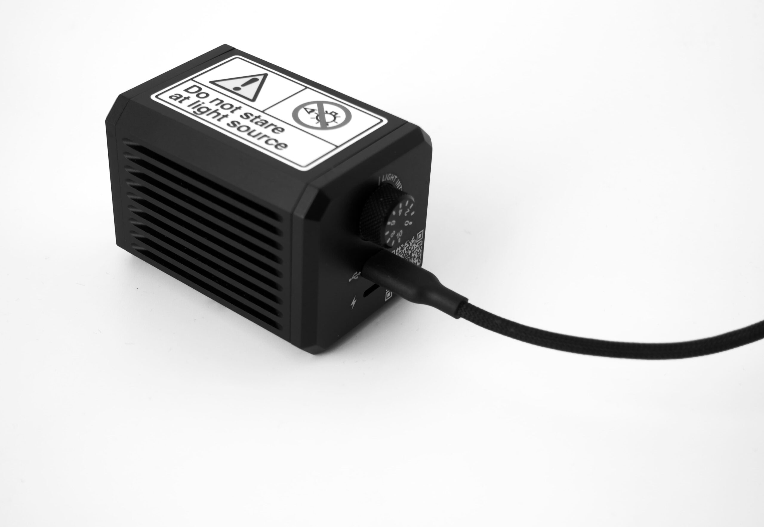
Near-Infrared Fluorescence Imager — an affordable and user‑friendly observation device for real-time tissue visualization.
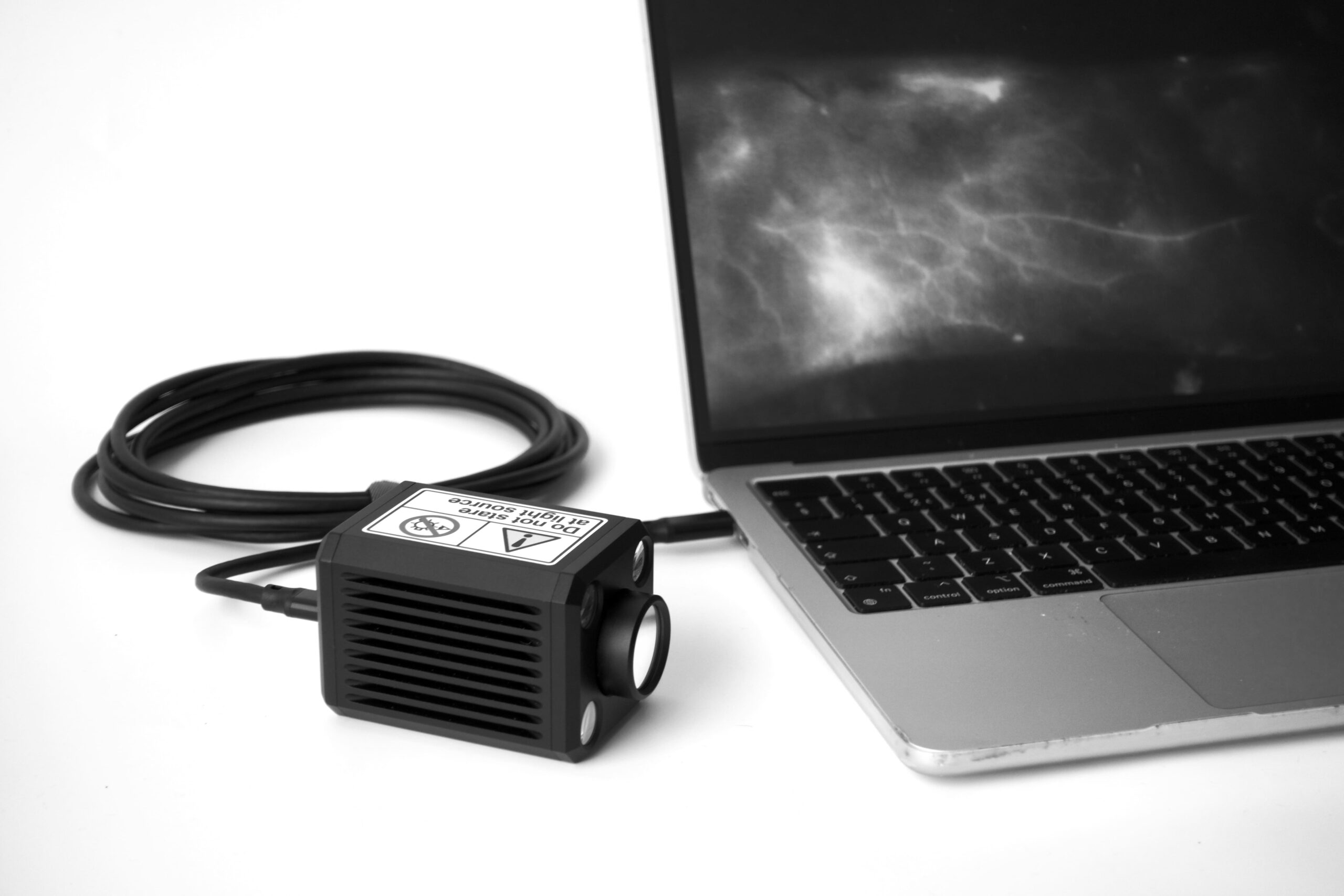
Near-Infrared Fluorescence Imager — an affordable and user‑friendly observation device for real-time tissue visualization.
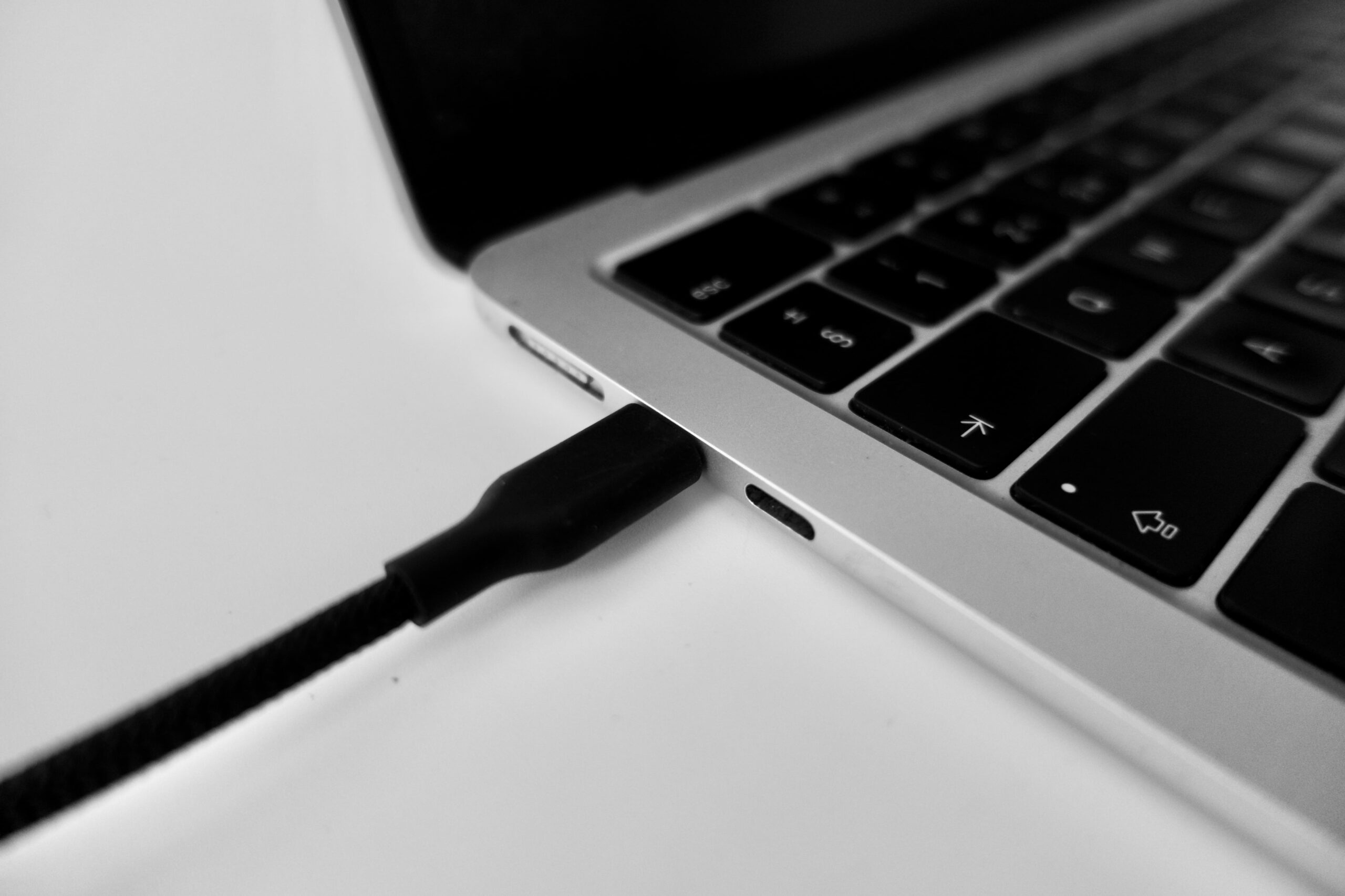
Simply connect to your PC via the included 3 m USB cable — extendable to approximately 5 m for flexible positioning.
Images save directly to your computer. No external DVD or Blu-ray recorder required — streamlining your workflow.
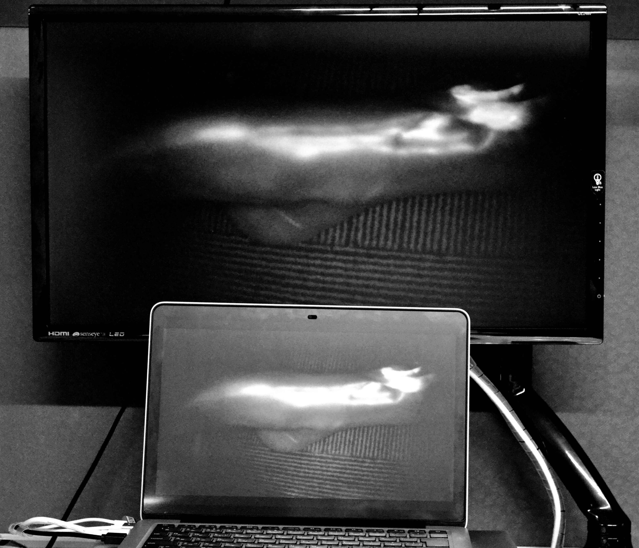
Output to an external monitor via your PC for large-screen observation during procedures or presentations.
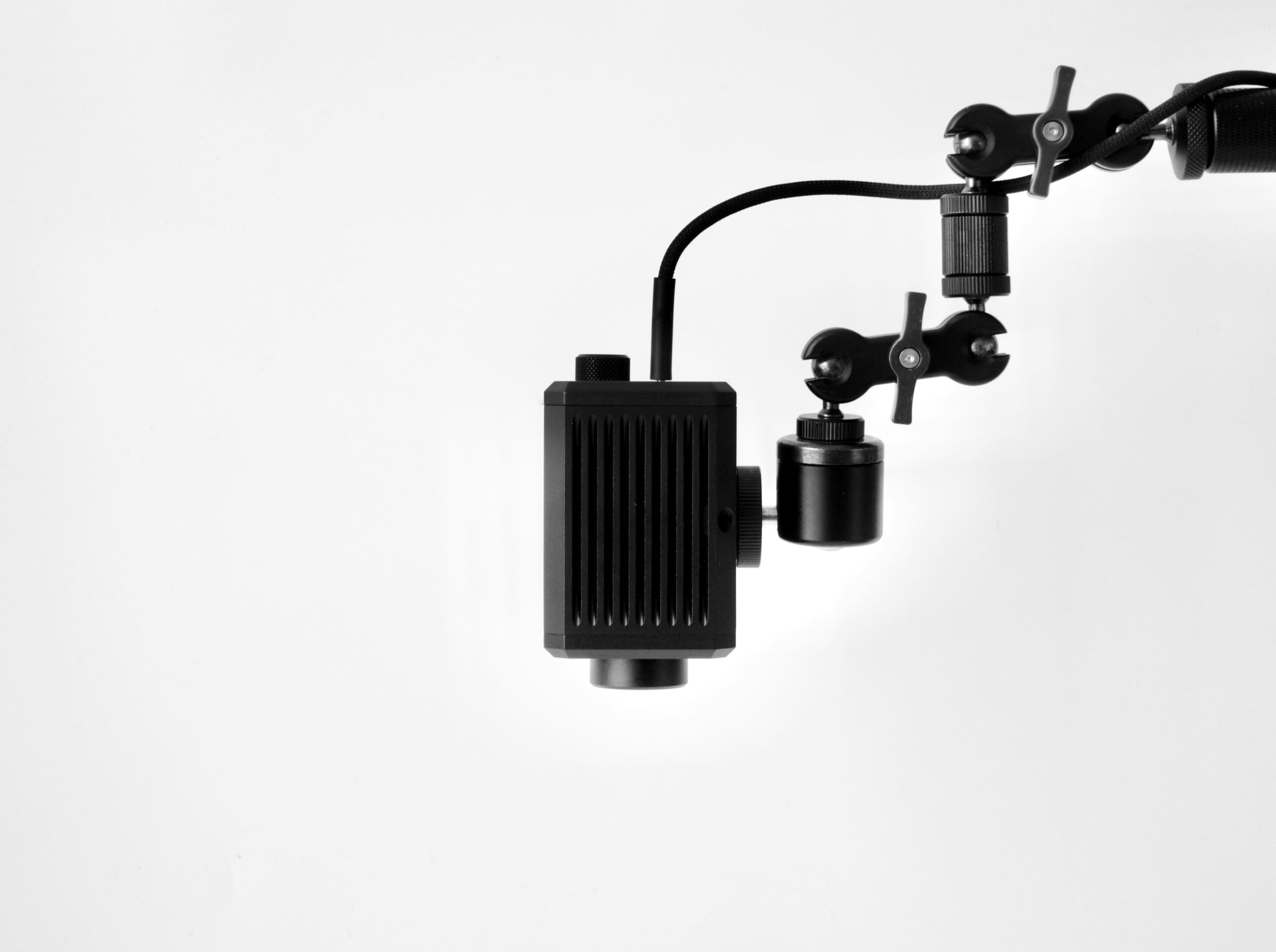
Mounts on any standard tripod for fixed-field observation, hands-free imaging, and consistent positioning.
Plug NIRFI™ into your PC using the included USB cable. The device is recognized by your PC with standard drivers — no complex setup required.
Mount on a tripod or handheld. Point at the area of interest. The 5 m cable reach gives you flexibility in any setting.
Capture real-time near-infrared fluorescence imagery. Images save instantly to your PC — ready for analysis or export.
Watch how NIRFI™ performs in real clinical and research scenarios.
Full technical details for integration planning and procurement.
| Parameter | Specification |
|---|---|
| Device Weight | Approx. 0.5 kg (device only) |
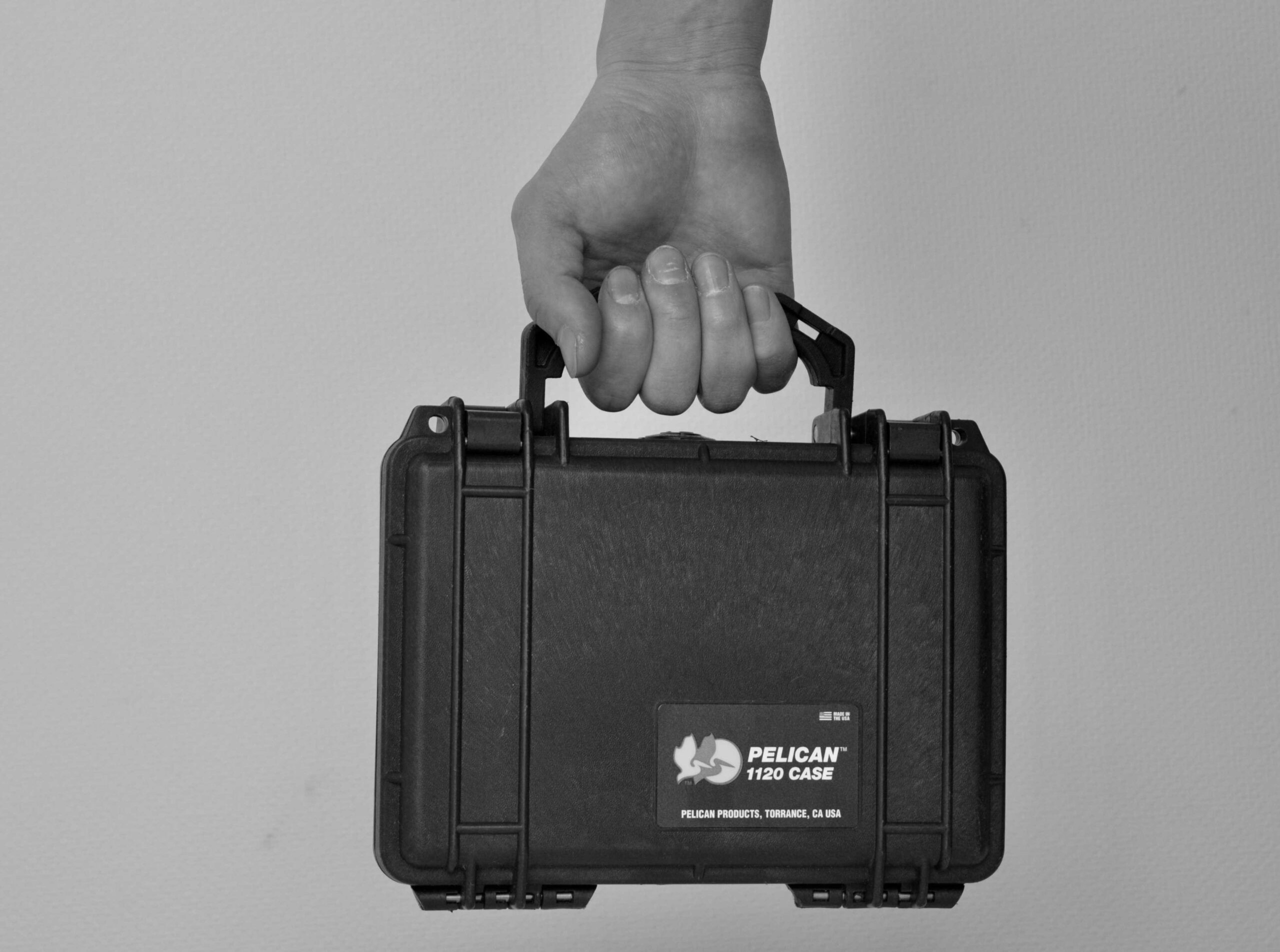
| Total Weight (with case) | Approx. 1.5 kg |
| Connectivity | USB |
| Cable Length | 3 m (included); extendable to approx. 5 m |
| Image Storage | Direct to PC — no external recorder required |
| Display Output | PC monitor; supports external monitor via PC output |
| Mounting | Standard tripod mount compatible |
| Imaging Modality | Near-infrared fluorescence (NIR) |
| Power Source | USB bus power |
| Compatible OS | Windows (details available on request) |
| Intended Use | Tissue observation and fluorescence imaging for research and professional use |
* Specifications subject to change without notice. Contact us for the latest datasheet.
Real-time visualization of tissue perfusion and lymphatic drainage during surgical procedures.
Lightweight enough to use at the bedside, in outpatient clinics, or in resource-limited environments.
Small animal imaging, drug delivery tracking, and fluorescence-guided tissue analysis.
Non-invasive blood flow evaluation using ICG or other NIR fluorescent agents.
Fluorescence-guided sentinel lymph node identification for oncological procedures.
Affordable enough to equip teaching labs and training programs with real NIR imaging capability.