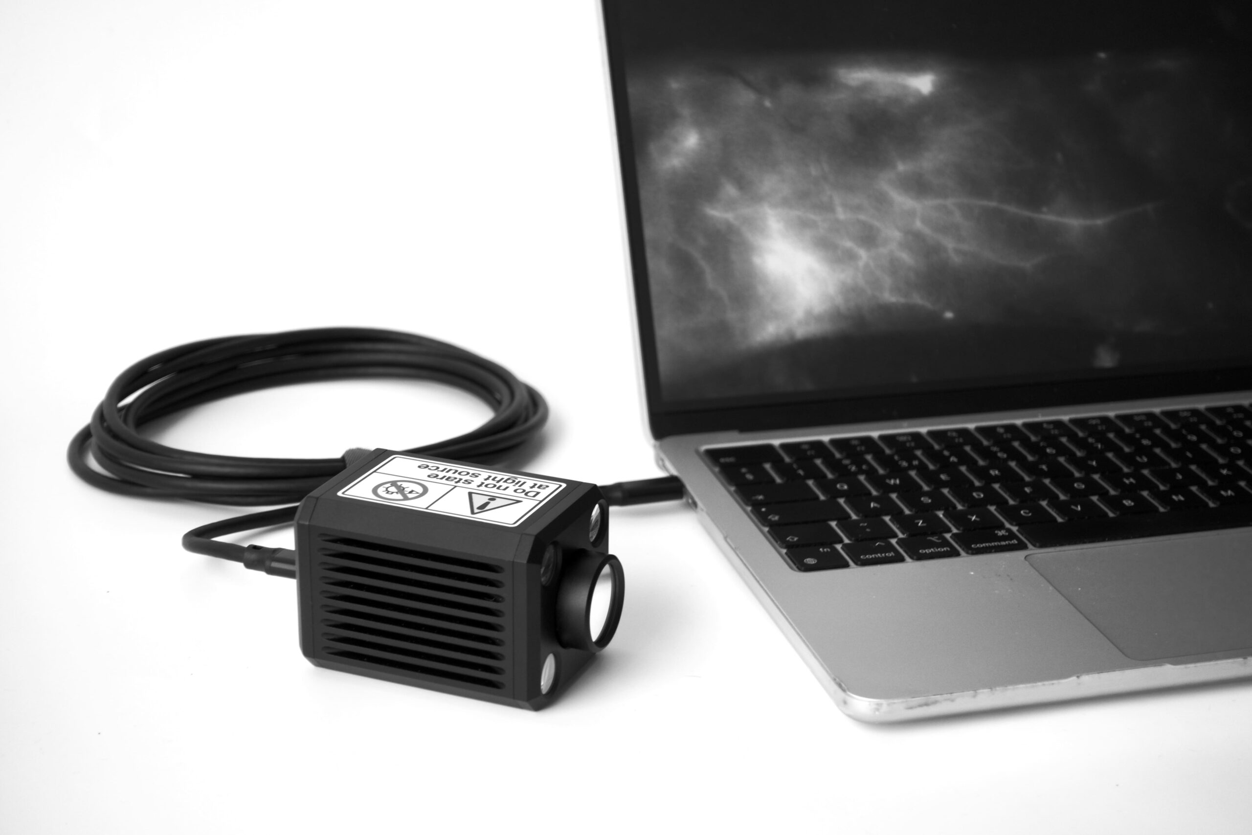
Near-Infrared Fluorescence Imaging
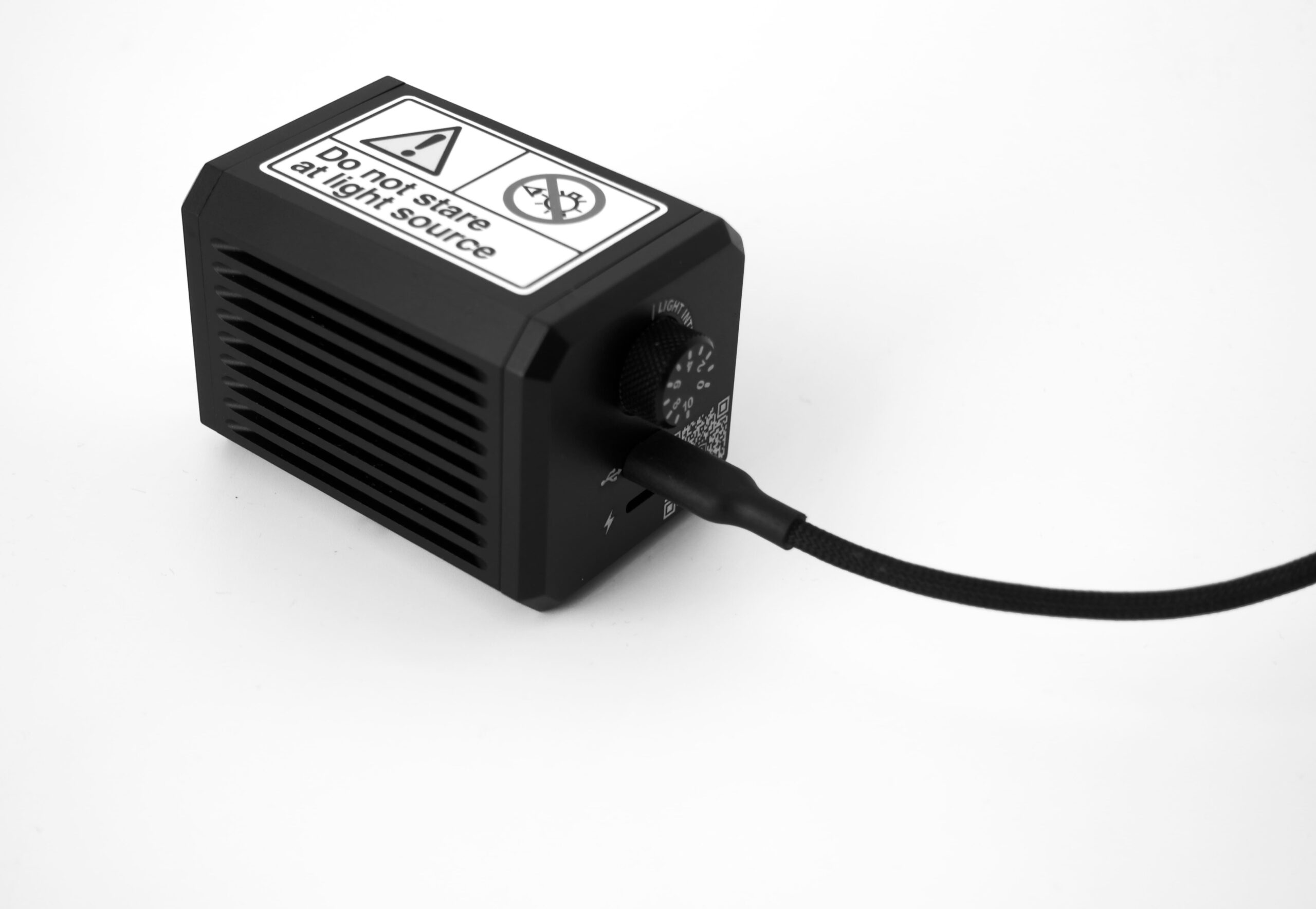
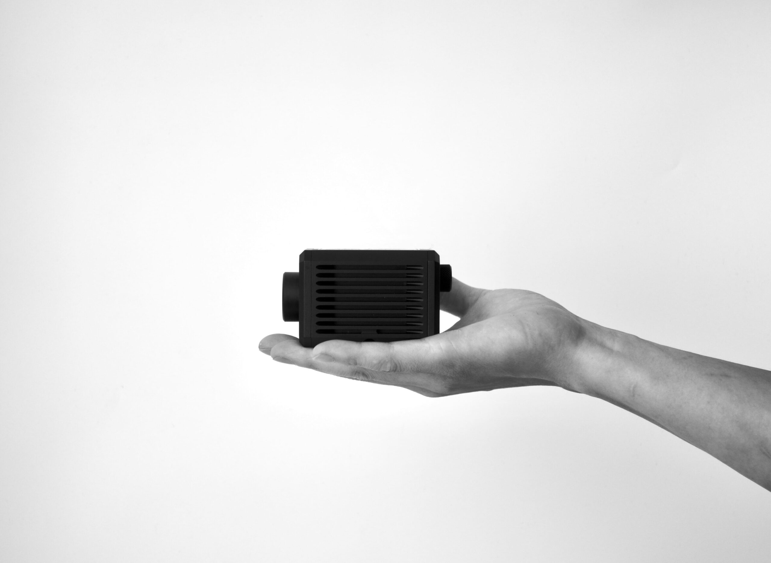
Compact.
Portable.
Powerful.
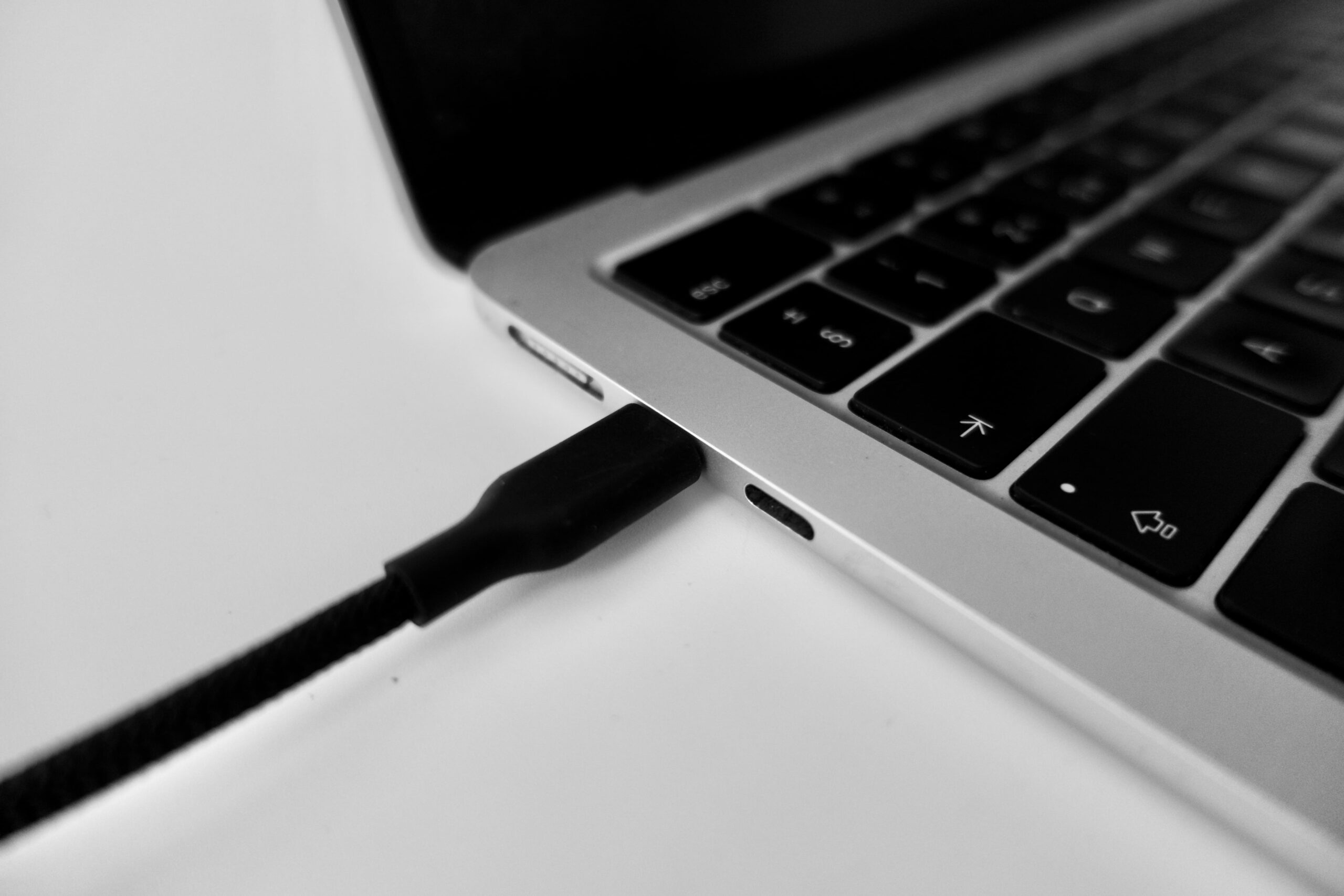
NIRFI™ enables near‑infrared fluorescence visualization of tissue for surgical guidance, research and training — now palm-sized.
Scroll